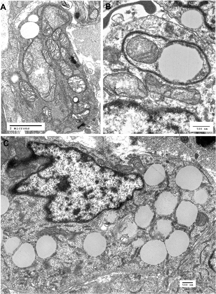
Figure. 2.
Ultrastructural features of the PDX cells. A. Electron micrograph of xenograft cell showing markedly abnormal mitochondria with swelling and loss of cristae. B. Xenograft cell with an autophagic vacuole containing a mitochondrion and amorphous material suggesting advanced degeneration of a mitochondrion or other cytoplasmic content. C. Xenograft cell containing numerous cytoplasmic vacuoles.

