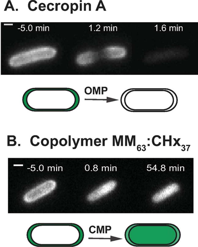
Figure 1.

Single-cell, time-lapse assay imaging periplasmic GFP to detect membrane permeabilization events induced in live E. coli by antimicrobial agents. (A) Cecropin A at 0.9 μM (1X MIC) first induces outer membrane permeabilization to GFP, beginning at the septal region. Initial periplasmic halo image gradually disappears. (B) Gellman random β-peptide copolymer MM63:CHx37 at 30 μg/mL (1.2X MIC) first induces cytoplasmic membrane permeabilization to GFP. Initial periplasmic halo image evolves to a cytoplasmic filled image of which persists for at least 55 min.
