
Figure 5.
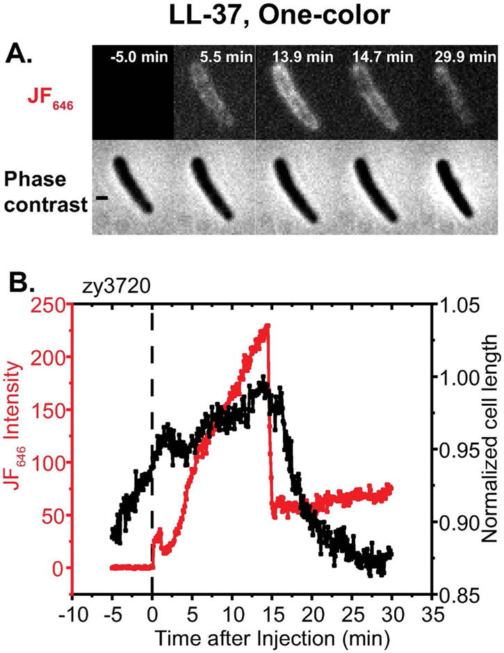
Application of one-color assay to membrane permeabilization by the antimicrobial peptide LL-37. Top: Red fluorescence and phase contrast snapshots of single zy3720 E. coli cell following addition of 4 μM LL-37 (1X MIC) plus 100 nM JF646 ligand at t = 0. Time resolution is 6 sec/cycle = 0.1 min/cycle. Bottom: Time dependence of cell length (from phase contrast images) and HaloTag-JF646 conjugate fluorescence intensity for the same cell. The fluorescence builds up in the periplasm for ~10 min, prior to the onset of significant cell shrinkage. Abrupt loss of some 75% of the intensity is attributed to OM permeabilization to globular proteins including GFP, HaloTag protein and HaloTag-JF646 conjugate. Simultaneous loss of GFP and HaloTag-JF646 conjugate was demonstrated in the two-color experiment of Figure S3. Scale bar is 1 μm.