
Figure 6.
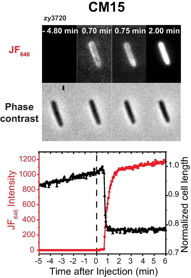
Application of one-color assay to membrane permeabilization by the antimicrobial peptide CM15. Top: Red fluorescence and phase contrast snapshots of single zy3720 E. coli cell following addition of 10 μM CM15 (2X MIC) plus 100 nM JF646 ligand at t = 0. Time resolution is 3 sec/cycle = 0.05 min/cycle. Bottom: Time dependence of cell length (from phase contrast images) and HaloTag-JF646 fluorescence intensity for the same cell. There is evidence of a transient periplasmic image at 0.70-0.75 min. The onset of red fluorescence and of cell shrinkage are simultaneous within one camera frame (3 sec).