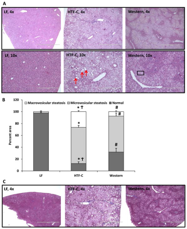
Figure 2.
The pattern and characteristic of hepatic steatosis differs between the HTF-C diet and Western diet fed mice. [A] Representative hematoxylin and eosin-stained liver sections from mice fed LF, HTF-C, and Western diet. [B] Percent macrovesicular steatosis versus microvesicular steatosis in liver specimens of mice fed LF, HTF-C, and Western diet. Red arrows indicate representative macrovesicular steatosis. Box indicates representative area of microvesicular steatosis. n=5 mice/group. [C] Representative Masson’s trichrome stain from mice fed LF, HTF-C, and Western diet fed mice. n=5 mice/group. Values are mean ± standard error of the mean. *p<0.05 LF vs HTF-C. #p<0.05 LF vs Western. †p<0.05 HTF-C vs Western.

