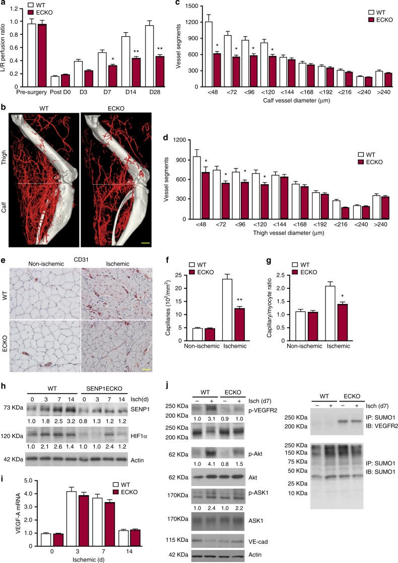
Fig. 1.
SENP1-ECKO mice exhibit attenuated arteriogenesis and angiogenesis in vivo. WT and SENP1-ECKO mice were subjected to hindlimb ischemic ligation model (HLI). a Laser Doppler analysis of blood flow. The graph shows blood flow in the ischemic foot expressed as a ratio to flow in the normal foot. Quantitative analysis of laser Doppler images indicates significant alterations in hindlimb reperfusion starting at 7 days after femoral artery ligation in SENP1-ECKO mice relative to WT mice (n = 6 each strain). b Representative micro-CT images of WT and SENP1-ECKO mice 14 days after HLI. c, d Quantitative micro-CT analysis of arterial vasculature above and below the knee in WT mice (n = 6500 cross-sections per mouse) and SENP1-ECKO mice (n = 4500 cross-sections per mouse) 14 days after common femoral artery ligation. Note a marked decrease in total number of <120-μm-diameter vessels in SENP1-ECKO mice relative to WT littermates in thigh and calf (mean ± SEM, *P < 0.05). Statistical significance was assessed using a Mann–Whitney U test and repeated measures analysis performed using one-way nonparametric ANOVA (Kruskal Wallis test). e–g Attenuated angiogenesis in SENP1-ECKO mice. Capillary density was immunostained with an EC marker CD31. Representative sections from non-ischemic and ischemic groups of WT and SENP1-ECKO mice on day 28 post-ischemia are shown in (e). Quantification of capillary density (number/mm2 muscle area) and ratio of CD31/myocyte are shown in (f, g). Data are mean ± SEM from ten fields per section (three sections/mouse and n = 4 for each strain). h–j Attenuated VEGF-VEGFR2 signalling in SENP1-ECKO. Muscle tissues from WT and SENP1-ECKO were harvested at various days post-ischemia as indicated. h HIF-1α, SENP1, and β-actin were determined by western blot with respective antibodies. i VEGF-A mRNA was measured by qRT-PCR with GAPDH for normalization. Fold changes are presented. n = 2 per group. j Phosphorylations of VEGFR2, Akt, and ASK1 as well as total proteins as indicated in tissue lysates were determined by western blot with respective antibodies. SUMOylated VEGFR2 was determined by co-immunoprecipitation assays with anti-SUMO1 followed by western blotting with anti-VEGFR2. Protein bands in (h, j) were quantified by densitometry and fold changes are presented by taking WT non-ischemia as 1.0. n = 2. Error bars, mean ± SEM; ∗P < 0.05, **P < 0.01, one-way ANOVA. Scale bar: 500 μm (b); 20 μm (e)

