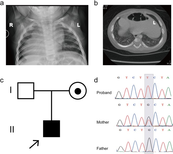
Figure 1.
Clinical features of the proband and the ZIC3 gene mutation. (a) Chest radiograph showing dextrocardia in the patient. (b) CT scan indicating that the liver and spleen of the patient are reversed, while the stomach is located in the middle of the body. (c) The trio family of the proband, in which the mother is a carrier. (d) Sanger sequencing results show that the hemizygous ZIC3 mutation was found in the proband, while his father did not carry this mutation, but his mother was a heterozygous carrier. The gray background indicates the nucleotide change from G to T in the patient at position 890.

