Figure 3.
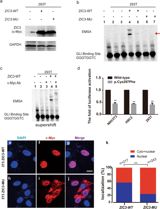
The ZIC3 mutation causes functional changes in cell lines. (a) A western blot showed that the ZIC3 wildtype (WT) and mutant (MU) forms are being expressed as equal levels for the EMSA experiment. (b) The ZIC3 domain binds to the GLI-binding site (GLBS). Line 1: Only the GLI probe without protein; line 2: GLI probe + protein in 293T cells; line 3: unlabeled competitor GLI probe + protein in 293T cells; line 4: GLI probe + protein in 293T cells transfected with pZIC3 (WT)-myc; line 5: unlabeled competitor GLI probe + protein in 293T cells transfected with pZIC3 (WT)-myc; line 6: GLI probe + protein in 293T cells transfected with pZIC3 (MU)-myc; line 7: unlabeled competitor GLI probe + protein in 293T cells transfected with pZIC3 (MU)-myc. The red arrow indicates the complex of GLIBS with the ZIC3 protein. (c) A supershift EMSA showed that c-Myc antibody could specific bind with whole cell lysate which transfected pZIC3-myc construct. Line 1–4 added biotin-labeled probe. (d) The wild-type (pZIC3-myc) or mutant (p.Cys297Phe) ZIC3 construct was co-transfected into NIH/3T3, H9C2, and HEK-293T cells with pGL3-SV40 firefly and pRL-TK Renilla luciferase reporters. Luciferase activities were measured 24 hours post-transfection. The mean fold activation relative to the wild-type is shown. The results represent the average luciferase activation across a minimum of three individual experiments. Standard errors are indicated by vertical lines. “**” Denotes statistical significance (P < 0.05) by two-tailed, unpaired Student’s t-tests assuming unequal variance. (e–j) Subcellular localization of ZIC3 determined by immunofluorescence in NIH3T3 cell lines. For each construct, anti-Myc (panels f,i) and DAPI (panels e,h) staining is shown individually and merged (panels g,j). The wild-type (WT) construct is located in the nucleus (panel f), but the C297A missense mutation construct is located in both the cytoplasm and nucleus (panel i). Scale bar indicates 63X magnification. (k) Percentage of localization. Cells transfected with the WT or MU ZIC3 construct were classified as exhibiting either only nuclear localization or both nuclear and cytoplasmic localization. ***P < 0.0001 by the Chi-square test.

