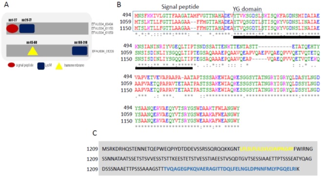
Figure 1.
Domain organization and alignment of E. faecium LysM domain–containing proteins. (A) Domain organization of the LysM-containing proteins. EFAU004_00494, EFAU004_01150 and EFAU004_01059 have the same organization, whereas EFAU004_01209 is organized differently. Red circle: signal peptide; blue square: LysM domain; yellow triangle: transmembrane domain. (B) Clustal Omega alignment of proteins EFAU004_00494, EFAU004_01150 and EFAU004_01059. The signal peptide and the conserved YG domain are boxed. The LysM domain is indicated by the black line below the alignment. (C) Sequence of the EFAU004_01209 LysM protein. The transmembrane helix and the LysM motif are highlighted in yellow and blue, respectively.

