Figure 1.
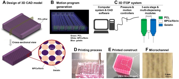
Bioprinting of skeletal muscle. (A) Design concept using 3D CAD modeling and (B) motion program generation of the bioprinted muscle construct. The code includes XYX stage movement and actuating pneumatic pressure. (C,D) Bioprinting process using ITOP system. (C) The motion program was transferred to the operating computer of ITOP. The cell-laden bioink containing hMPCs, the acellular sacrificing hydrogel, and the supporting PCL pillar were loaded in the multi-dispensing modules. (D) All three components were printed in a layer-by-layer fashion. (E) The bioprinted skeletal muscle constructs composed of multi-layered myofiber bundles were fabricated up to 15 × 15 × 15 mm3 in dimension. The thickness of the printed muscle construct was determined by controlling the number of stacking myofiber bundles. (F) Microchannels in the constructs created after the removal of the sacrificial patterns to maintain the viability of printed cells.

