Figure 2.
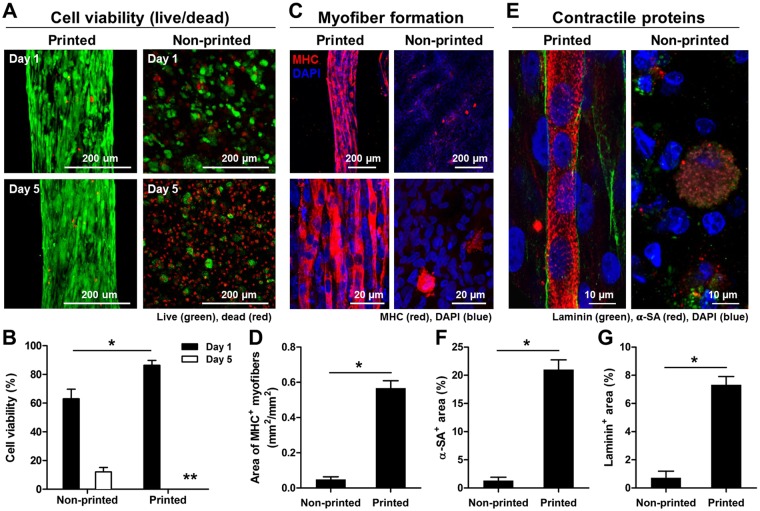
In vitro evaluations of bioprinted muscle constructs compared with non-printed constructs. (A) Representative Live/Dead staining images and (B) cell viability (%) at 1 and 5 days in culture (n = 4, 4 random fields/sample, *P < 0.05, **not measurable because of too confluence - % viability was over 90%). (C) Immunofluorescent staining for MHC after 7 day-differentiation and (D) quantification of area of MHC + myofibers (n = 3, 4–7 random images/sample, *P < 0.05). Human MPCs in the construct showed enhanced myofiber formation with unidirectional cell alignment. (E) Double-immunostaining for α-SA (red)/laminin (green) indicates the presence of cross-striated myofibers surrounded by laminin matrix in the printed construct. Quantification of (F) α-SA+ area (%) and (G) laminin+ area (%) (n = 3, *P < 0.05).

