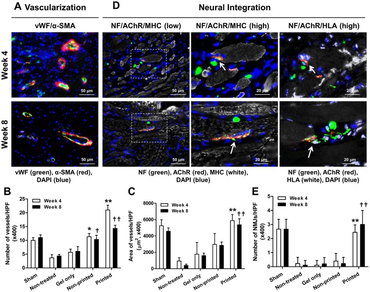
Figure 9.
Immunofluorescence of vascularization and neural integration of the implanted bioprinted muscle constructs. (A) Immunofluorescent images of vWF (green)/α-SMA (red) of the regenerated TA muscles at 4 and 8 weeks after implantation. Quantification of (B) vessels/field and (C) area of vessels/field (µm2) (n = 3, *P < 0.05 compared with non-treated and gel only groups, **P < 0.05 compared with other groups). (D) Immunofluorescent images of NF (green)/AChR (red)/MHC (white) and NF (green)/AChR (red)/HLA (white) of the regenerated TA muscles at 4 and 8 weeks after implantation. NF+/AChR+/MHC+ neuromuscular junction (middle column, white arrow) was observed in bioprinted muscle constructs. NF+/AChR+/HLA+ neuromuscular junction (right column) corresponding area NF+/AChR+/MHC+ (middle column) indicates that bioprinted muscle constructs are integrated with host nervous system following implantation. The white arrow indicates neuromuscular junction on hMPC-myofibers. (E) Quantification of NMJ/field (×400) (n = 3 per group, 3 random fields per sample, *P < 0.05 compared with other groups).

