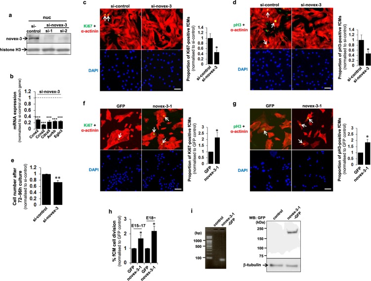
Figure 5.
Nuclear novex-3 is a cell cycle promoter in hypoxic fCMs. (a) Novex-3 was silenced in E17 fCMs by siRNA, and the nuclear fraction was used to probe novex-3 protein by immunoblotting. Two siRNAs specific for novex-3, designated as si-1 and si-2, were used. Histone H3 was included as a loading control. (b) qPCR analysis of mRNA levels of the indicated genes in novex-3-silenced fCMs (E16–E17). Data are shown as normalised to the si-control level of each gene, set at 1. n = 3 independent experiments. ***P < 0.001 compared to the si-control level of each gene. (c,d) Immunofluorescence for Ki67 (c, green) and phospho-histone H3 (pH3) (d), green) observed in sarcomeric α-actinin (red, as a CM marker) and DAPI (blue) in novex-3-silenced fCMs (E17). Arrows denote Ki67-positive (c) and pH3-positive (d) fCMs. Proportions of Ki67-positive (c) and pH3-positive (d) fCMs were shown on the right. n = 4 independent experiments. *P < 0.05 compared to si-control. Scale bars = 50 µm. (e) Proliferative activity of novex-3-silenced E16–E17 fCMs evaluated by cell counting after 72–96 h culture. n = 3 independent experiments. **P < 0.01 compared to si-control. (f,g) Immunofluorescence for Ki67 (f), green) and phospho-histone H3 (pH3) (g, green) observed in sarcomeric α-actinin (red, as a CM marker) and DAPI (blue) in novex-3-1-overexpressing fCMs (E15–E17). Arrows denote Ki67-positive (f) and pH3-positive (g) fCMs. Proportions of Ki67-positive (f) and pH3-positive (g) fCMs are shown at right. n = 4 independent experiments. *P < 0.05 compared to control GFP-overexpressing fCMs. Scale bars = 50 µm. (h) Percentage of novex-3-1-overexpressing fCMs that completed cell division, as determined by time-lapse imaging. Analysis was performed separately in two developmental stages (E15–E17 and later than E18). n = 3 independent experiments. *P < 0.05 compared to control GFP-overexpressing fCMs. (i) Left, overexpression of novex-3-1-GFP mRNA in E17 fCMs was confirmed by probing 61 bp amplicon within GFP sequence by PCR. Right, overexpression of novex-3-1-GFP protein in E17 fCMs was confirmed by immunoblotting the whole protein extracts using the antibody against GFP. A clear single band was observed at the expected molecular mass (novex-3-1: ~175 kDa + GFP: ~27 kDa). β-tubulin was included as a loading control. Error bars = SEM.

