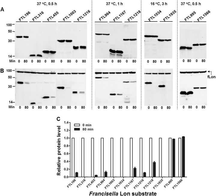
Fig. 1. Degradation of the F. tularensis proteins by fLon in E. coli ER2566 (Lon−).
Stability of the F. tularensis proteins in the absence (a) or presence (b) of the fLon-expression plasmid in Lon-deficient E. coli ER2566. fLon was induced with arabinose for 2 h before induction of the target proteins with IPTG under the conditions specified at the top of each panel, and subsequently treatment with spectinomycin. The cells were harvested at 0 and 80 min after the addition of spectinomycin; each target protein detected by immunoblotting using the anti-His6 antibody. The sizes of protein standards are indicated at the left side in kDa. c The amount of each target protein in b was quantified by Image Lab. The level of each protein at 80 min is presented as a value relative to that at 0 min. Bars represent the mean value ± SEM (n = 3)

