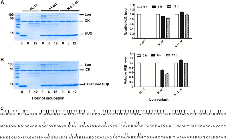
Fig. 7. Differential degradation of HUβ by E. coli and human Lon variants.
Recombinant HUβ (15 µg) was incubated at 37 °C with the Lon protease (10 µg) of E. coli (eLon) or human (hLon) before (a) or after (b) being denatured by heat. The proteins in the reactions were detected by SDS-PAGE and Coomassie Brilliant staining at 0, 6, and 12 h. Creatine kinase (CK) presented in the reaction mixture was used for ATP regeneration in this assay. HUβ was quantified by Image Lab and presented as relative value to the sample taken at 0 h (left panel of a and b). Sites of peptide bond break in HUβ in the presence of eLon (top panel) or hLon (middle), or in the absence of Lon protease (bottom) were identified by detection of peptides in the samples taken at 6 h by mass spectrometry (c). The arrows above the gaps of adjacent amino acids indicate the ends of individual peptides

