
Figure 1.
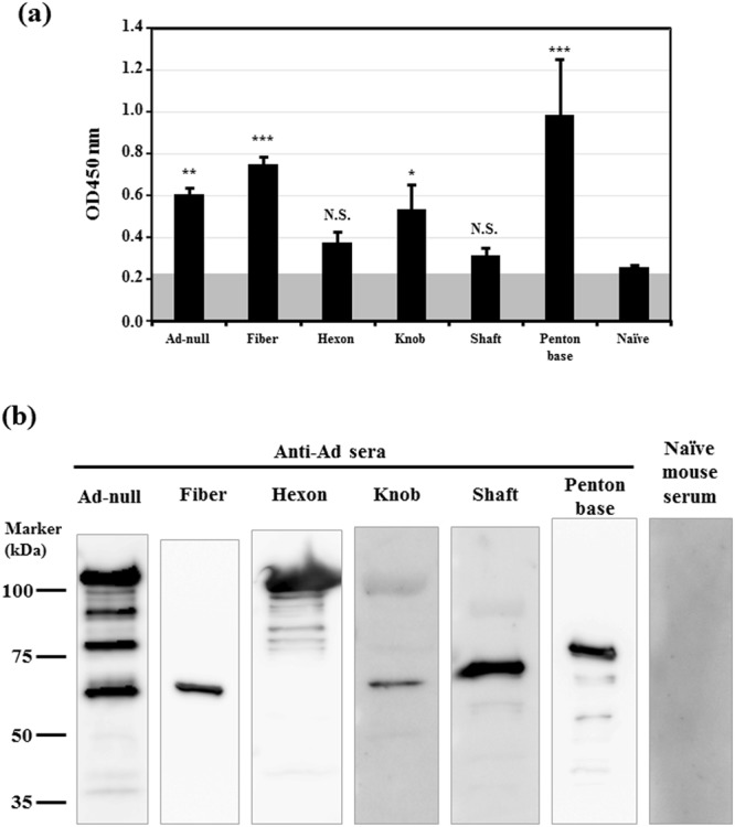
Analysis of Ad capsid protein-specific sera isolated from Ad capsid protein-expressing plasmid-immunized mice. (A) ELISA analysis using Ad capsid protein-specific sera. Ad-null was solubilized by 0.1% Triton X-100 and immobilized on a plate. Anti-Ad capsid protein sera were diluted and added to the well. The gray shaded boxes indicate background levels. The data are expressed as the mean ± S.D. (n = 4). n.s., not significant, *p < 0.05, **p < 0.01, ***p < 0.001, compared with naïve sera. (B) western blotting analysis using Ad capsid protein-specific sera. Ad-null was denatured at 98 °C for 5 min and loaded on SDS-PAGE gels according to the manufacturer’s protocol. Western blot analysis was carried out using anti-Ad capsid protein sera. Each image was captured under different exposure times. Representative images from three independent experiments using different mouse serum batches are shown.