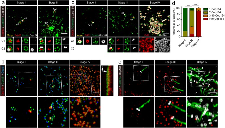
Figure 3.
Centriole maturation in MEFs expressing Multicilin and E2f4VP16. (a) Super-resolution images of MEFs expressing Multicilin/E2f4VP16 stained for Cep63 (red), Centrin (green) and Cep164 (gray) at different stages of MCC differentiation. Magnified insets show Cep164 staining is first associated with the pre-existing mother centriole (stage II), followed by the daughter centriole (stage III), both marked by Cep63 (C1, C2). (b) Super-resolution images of MEFs expressing Multicilin/E2f4VP16, stained for Cep164 (red), Centrin (green) and Deup1 (blue) at different stages of MCC differentiation. The magnified insets show the sequential appearance of Cep164 at the mother centriole, daughter centriole, and finally new centrioles as Deup1 staining disappears. The top and side view of Stage IV with Cep164 (red) and Centrin (green) shows centriole alignment and docking to the surface membrane. (c) Super-resolution images of MEFs expressing Multicilin/E2f4VP16, stained for Odf2 (red), Centrin (green) and Cep164 (gray) at different stages of MCC differentiation. The magnified insets show the acquisition of distal appendages (Cep164) and subdistal appendages (Odf2) are closely linked. (d) Fraction of infected MEFs containing different extents of Cep164 staining was scored at different stages of MCC differentiation. Only ring-shaped Cep164 staining pattern was counted, scoring >45 cells obtained in triplicate, at each stage. Error bars = s.d. (e) Super-resolution images of MEFs expressing Multicilin/E2f4VP16, stained with Deup1 (red), the cilia marker GT335 (green) and Cep164 (gray) at different stages. The magnified insets show that the acquisition of appendages precedes cilia projection. Scale bars = 2 μm.

