Figure 4.
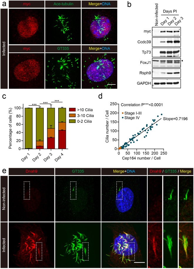
Cilia formation in MEFs expressing Multicilin and E2f4VP16. (a) Confocal images of MEFs expressing Multicilin/E2f4VP16, fixed and stained for the myc-tag on E2f4VP16 (red), the cilia markers, acetylated-Tubulin and GT335 (green), followed by DAPI staining (blue). (b) Western blot analysis of non-infected MEFs, or MEFs expressing Multicilin/E2f4VP16 with antibodies to myc, Foxj1, Tp73, Ccdc39 or Rsph9, at the indicated day PI. Arrowhead indicates non-specific band. (c) The fraction of MEFs expressing Multicilin/E2f4VP16 with the indicated number of cilia based on GT355 staining at the indicated day PI. Error bars = s.d. (d) Plot of cilia number per cell versus Cep164 number per cell, taking data from MEFs expressing Multicilin/E2f4VP16 at 1–4 days PI. Cep164 number (matured basal bodies) was scored based on ring-shaped Cep164 detected using super-resolution microscopy and cilia number by Arl13b staining. MEFs classified as stage I-III (as described in the text, orange points) contained few matured centrioles (just the MCD pathway), while those classified as stage IV (dark points) showed matured centrioles based on Cep164 staining that dock and form cilia. Pearson correlation analysis showed a strong correlation between centriole maturation and ciliogenesis with an average slope of 0.7196 indicating that acquisition of appendages precedes ciliogenesis. (e) Confocal images of MEFs, either non-infected or expressing Multicilin/E2f4VP16, stained for dynein heavy chain 9 (Dnah9, red), GT335 (cilia marker, green) and DAPI (blue). Magnified insets show MEFs expressing Multicilin/E2f4VP16 extend motile cilia. Scale bars = 10 μm.

