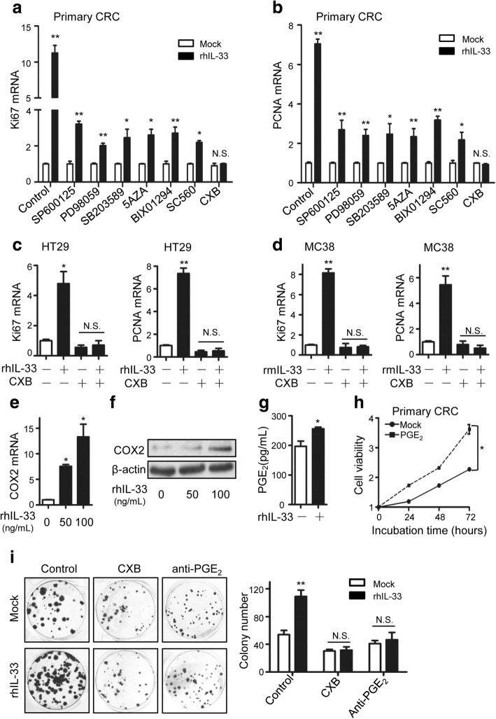
Fig. 2.
COX2/PGE2 mediates the proliferation promoting function of IL-33. a, b The relative mRNA levels of Ki67 (a) and PCNA (b) in primary CRC cells responding to rhIL-33 (100 ng/mL) incubation and/ or indicated inhibitors (SB203580, 10 μg/mL; PD98059, 20 μg/mL; SP600125, 10 μg/mL; BIX01294, 2 μM; 5Aza, 10 μM; SC560, 0.1 μg/mL; celecoxib, 20 μg/mL) for 24 h. c The relative mRNA levels of Ki67 and PCNA in HT-29 cells incubated with rhIL-33 (100 ng/mL) or/ and celecoxib (CXB) (20 μg/mL) in medium for 24 h. d The relative mRNA levels of Ki67 and PCNA in MC38 cells incubated with rmIL-33 (100 ng/mL) or/ and celecoxib (CXB) (20 μg/mL) in medium for 24 h. e, f The mRNA (e) and protein (f) expression of COX2 in primary CRC cells incubated with 0, 50 or 100 ng/mL of rhIL-33 in medium for 24 h. g PGE2 concentrations in the supernatants of primary CRC cells incubated with rhIL-33-contained RPMI medium or blank RPMI medium for 48 h. h Cell viabilities of primary CRC cells incubated with or without PGE2 (50 ng/mL) in medium. i The flat colony formation of primary CRC cells incubated for 15 days in medium containing different factors as indicated (IL-33, 100 ng/mL; celecoxib, 20 μg/mL; anti-PGE2, 2 μg/mL). The representative images of colonies and the statistical data are shown. Three parallel wells were set for each treatment. Each experiment was performed three times. Data expressed as mean ± SEM. * P < 0.05. ** P < 0.01

