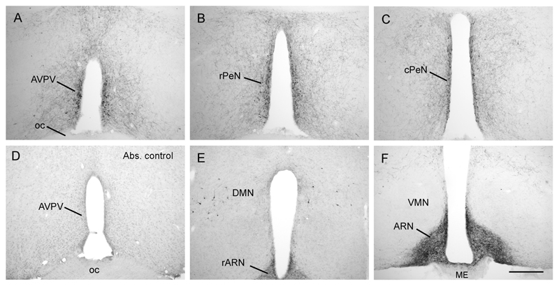
Figure 1. Distribution of kisspeptin immunoreactivity in the mouse hypothalamus.
Kisspeptin cell bodies were found in a periventricular continuum beginning in the anteroventral periventricular nucleus (AVPV; A) and extending caudally through into the preoptic periventricular nucleus (PeN), divided here into the rostral (rPeN; B) and caudal (cPeN; C) halves for analyses. There was a complete absence of labelling in tissue that was incubated with kisspeptin-10 antisera that was preadsorbed with mouse kisspeptin-10 peptide (D). Kisspeptin cell bodies were also present scattered within the dorsomedial nucleus (DMN; E), while dense fiber staining filled the arcuate nucleus (ARN; F) with cell bodies only occasionally discernable. ME, median eminence; oc, optic chiasm; VMN, ventromedial nucleus. Scale bar in F is 300μm.

