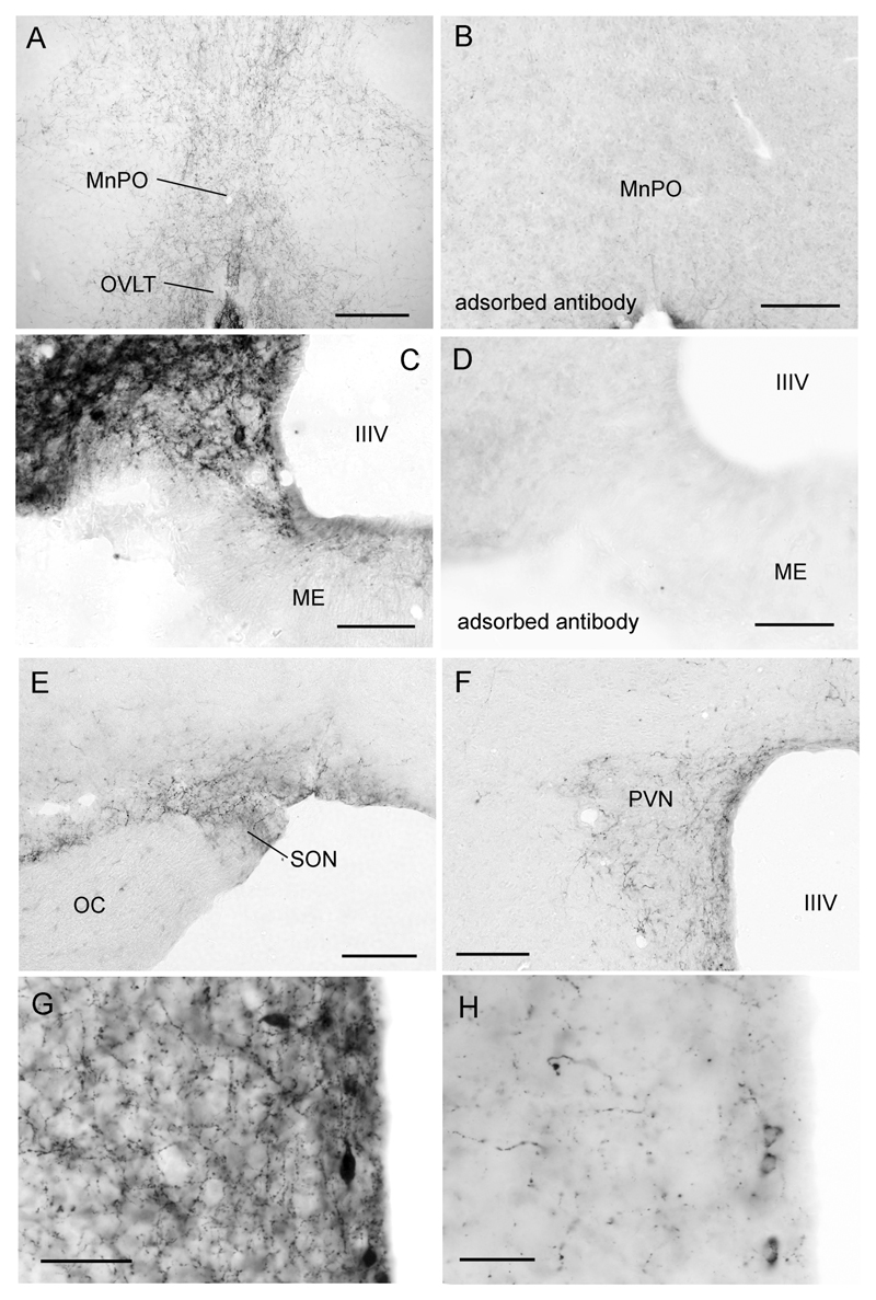
Figure 2. Distribution of kisspeptin immunoreactive fibers in the mouse hypothalamus.
A low-power view of kisspeptin fibers within the rostral preoptic area where the largest density of GnRH neuron cell bodies are found. B same region but following staining with kisspeptin antiserum adsorbed with kisspeptin-10. C Higher-power view of kisspeptin fiber plexus within the arcuate nucleus (ARN). Note the cell body profile at the ventral edge of the ARN and lack of staining in the external zone of the median eminence (ME). D same region but following staining with kisspeptin antiserum adsorbed with kisspeptin-10. Kisspeptin fiber staining was also found in the supraoptic (E) and paraventricular (F) nuclei. G and H show high-power images of kisspeptin-immunoreactive fiber and cell bodies in the preoptic periventricular nucleus in postnatal day 61 (P61) and P25 female mice, respectively. Abbreviations = MnPO, median preoptic nucleus; OVLT, organum vasculosum of the lamina terminalis; IIIV, third ventricle; ME, median eminence; OC, optic chiasm; SON, supraoptic nucleus; PVN, paraventricular nucleus. Scale bars represent 240μm (A,B,E,F) and 120μm (C,D,G,H).

