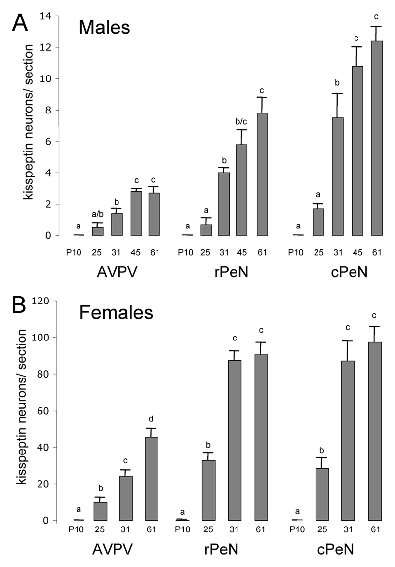
Figure 5. Quantitative analysis of kisspeptin-immunoreactive cell bodies in the developing periventricular nuclei.
Data are shown for the anteroventral periventricular (AVPV) and preoptic periventricular nucleus (PeN) divided into rostral (rPeN) and caudal (cPeN) halves. The mean (+SEM) number of immunoreactive cells detected per section at the three levels and at the indicated postnatal days (P) is given for males (A) and females (B). Bars labeled with different letters are significantly different from each other at either p<0.05 or p<0.01 (see text). N= 4-8 for each sex and age group.

