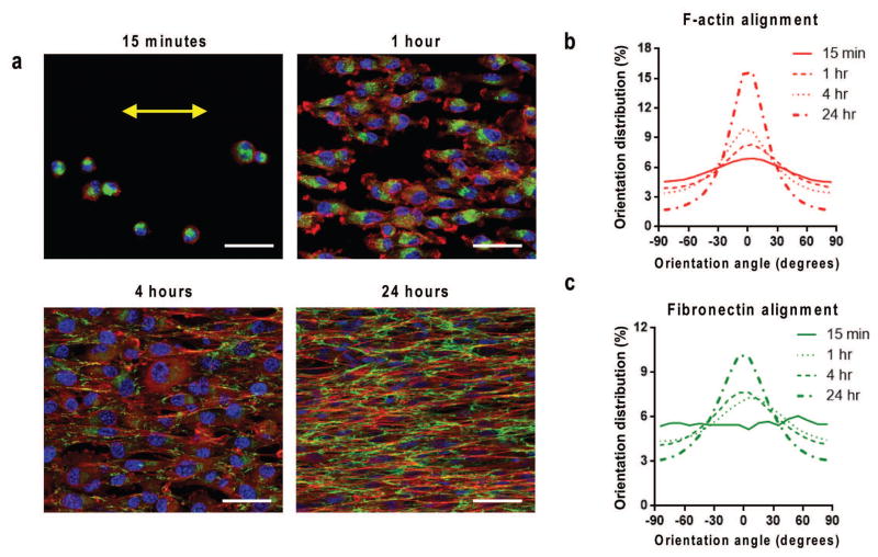
Figure 2. Rapid anisotropic cell attachment, cytoskeletal and ECM alignment.
(a) Confocal microscope image of F-actin (red), fibronectin (green), and nuclei (blue) showing TNFS-seeded C2C12 myoblasts at varied time intervals following seeding on nanotopographical substrates, demonstrating the rapid detection and alignment due to nanotopographical cues. Time course quantitative analysis of (b) cytoskeletal alignment and (c) cell-deposited ECM alignment of cells relative to major axis. Substrate nanopattern orientation is along the horizontal axis. Scale bars: 50μm.

