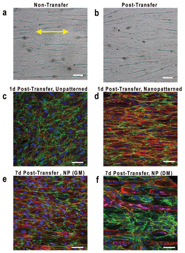
Figure 3. Myoblasts cultured on nanopatterned substrates align and secrete ECM in response to nanotopographical cues.
Bright field microscope images of myoblast monolayers differentiated for 7 days while (a) on TNFS or (b) transferred to a flat glass surface with formation of aligned myotubes. Yellow double-sided arrows indicate initial substrate nanopattern direction. Scale bar, 300 um. Confocal microscope images of F-actin (red), fibronectin (green), and nuclei (blue) staining showing gel-casted and transferred (c) unpatternend and (d) nanopatterned myoblast bilayer sheets 24 hours after transfer onto a flat glass surface. Confocal microscope images of nanopatterned bilayer sheets including MY-32 (magenta) following 7 days in (e) quiescent growth conditions and (f) differentiation conditions. Substrate nanopattern orientation is along the horizontal axis. Scale bars: 50 um.

