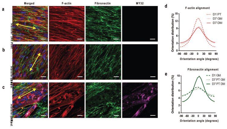
Figure 4. Transferred nanopatterned myoblast sheets remodel cytoskeletal and ECM structure depending on culture condition.
Confocal microscope image of F-actin(red), fibronectin (green), MY-32 (magenta) and nuclei (blue) staining to visualize structural organization of transferred myoblasts under (b) growth conditions and (c) differentiation conditions. Sheets cultured under growth conditions did not stain positive for myotubes. Yellow double-sided arrows indicate initial substrate nanopattern direction. Quantitative analysis of distribution of (d) cytoskeletal alignment and (e) cell-deposited ECM alignment under varied culture conditions of transferred nanopatterned cell sheets relative to major axis. Scale bars: 20μm.

