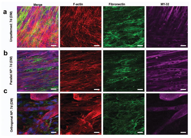
Figure 5. Stacked myoblast bilayers maintain cytoskeletal and ECM structure long term under quiescent conditions.
Confocal microscope image of F-actin (red), fibronectin (green), MY-32 (magenta) and nuclei (blue) staining to visualize structural organization of transferred myoblasts under differentiation conditions 7 days after transfer of (a) unpatterned, (b) parallel, and (c) orthogonal bilayers. Scale bars: 20μm.

