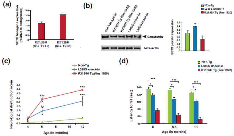
Figure 1. Characterization of expression and motor function of SETX ALS4 mice.
(a) Human SETX transgene expression levels from whole brain RNAs were determined by Taqman qRT-PCR analysis for two month-old SETX-R2136H mice (lines #1917 and #1920). Expression level is shown relative to endogenous murine Setx, and normalized to Gapdh. n = 3 mice/genotype, n = 3 technical replicates.
(b) SETX immunoblot analysis of brainstem protein lysates for two-month old mice of the indicated genotype. Beta-actin served as the loading control. Densitometry quantification of SETX normalized to beta-actin is shown in the graph to the right, with non-transgenic (Non-Tg) arbitrarily set to 1.
(c) Composite neurological phenotype analysis of mice of the indicated age and genotype (n = 6 – 8 mice/genotype). **P < .01, ***P < .001; ANOVA with post-hoc Bonferroni test.
(d) We measured the average latency to fall from the accelerating rotarod for mice of the indicated age and genotype (n = 8 mice/genotype, 3 runs/day averaged over 4 days). The same cohorts of mice were used for the entire experiment. *P < .05, ***P < .001; ANOVA with post-hoc Bonferroni test. Error bars = s.e.m.

