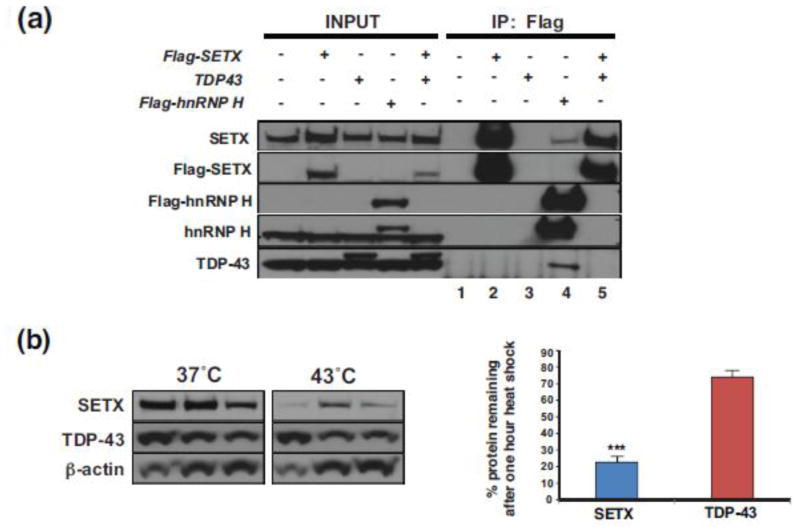
Figure 5. SETX protein does not interact with TDP-43 and is less stable than TDP-43.
(a) HEK293A cells were transfected with a Flag-SETX expression vector, Myc-TDP-43-HA expression vector, and/or a Flag-hnRNP H expression vector as indicated, and then subjected to immunoprecipitation (IP) with an anti-Flag antibody. Immunoblot analysis with the indicated antibodies was performed. Note that anti-TDP-43 immunoblotting of Flag-SETX IPs in lanes 2 and 5 does not detect TDP-43, but immunoblotting of the Flag hnRNP H IP (lane 4) detects both SETX and TDP-43, confirming the validity of the IP technique. No detectable proteins in lanes 1 and 3 rule out artefacts of the Flag IP technique, as these samples did not contain any Flag-tagged proteins.
(b) HEK293A cells were maintained at 37°C or raised to the heat shock temperature of 43°C for 1 hr. Immunoblot analysis of protein extracts after one hour at 37°C or 43°C (n = 3 separate cell cultures/treatment, as shown) indicate limited turnover of TDP-43, but marked reduction in SETX protein at the heat shock temperature of 43°C. Beta-actin served as the loading control. Densitometry quantification of SETX and TDP-43 protein levels normalized to beta-actin is shown in the graph to the right. ***P < .001, t-test. Error bars = s.e.m.

