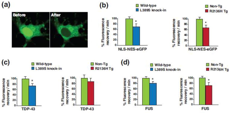
Figure 8. SETX ALS4 mice display defective nucleocytoplasmic transport.
(a) Primary cortical neurons from Non-Tg, PrP-SETX-R2136H, and SETX-L389S+/− mice were transduced with lentivirus containing a NLS-NES-eGFP shuttling reporter and subjected to photobleaching of the nucleus. Here we see the distribution of eGFP fluorescence in transduced cortical neurons before and after photobleaching of the nucleus.
(b) We performed FRAP of NLS-NES-eGFP transduced primary cortical neurons from mice of the indicated genotypes, and measured the recovery of eGFP signal in the nucleus over time (n = 4 mice/genotype). Fluorescence recovery/min was set to 100% for Wild-type or Non-Tg. *P < .05, t-test.
(c) We performed FRAP of TDP-43-mGFP transduced primary cortical neurons from mice of the indicated genotypes, and measured the recovery of mGFP signal in the nucleus over time (n = 3 mice/genotype). Fluorescence recovery/min was set to 100% for Wild-type or Non-Tg. *P < .05, t-test.
(d) We performed FRAP of FUS-mGFP transduced primary cortical neurons from mice of the indicated genotypes, and measured the recovery of mGFP signal in the nucleus over time (n = 3 mice/genotype). Fluorescence recovery/min was set to 100% for Wild-type or Non-Tg. *P < .05, t-test. Error bars = s.e.m.

