
Figure 3. NLRP3 expression in human testes is associated with phenotypic characteristics in subfertility patients.
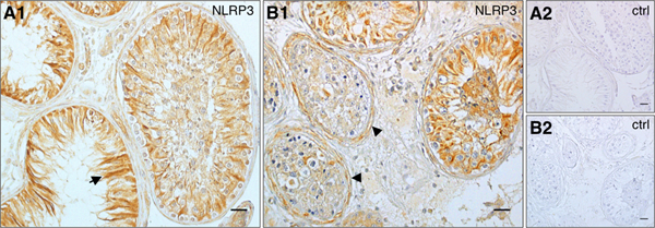
(A1) In patients suffering from mixed atrophy (MA) syndrome (n = 5) staining in Sertoli cells remained prominent, especially in tubules with impaired spermatogenesis (arrow). (B1) Staining of peritubular cells and the tubular wall (arrowheads) intensified corresponding to thickened sectors of the tubular wall, which are associated with MA pathology. Tubular walls stained most intensely in seminiferous tubules, which lacked Sertoli cell staining. (A2, B2) Negative controls corresponding to A1 and B1, respectively. Bars = 25 μm.