Fig. 1.
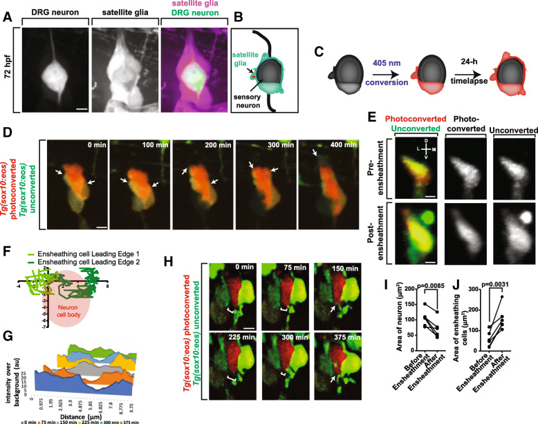
Sensory neurons become ensheathed shortly after neuronal differentiation. (a). Confocal z-projection frame of a Tg(sox10:mcherry); Tg(ngn1:gfp) zebrafish DRG at 72 hpf showing complete ensheathment. (b). Diagram of sensory neuron cell soma ensheathment by satellite glia. (c). Diagram of Eos photoconversion paradigm before neuronal differentiation. (d). Confocal z-projection images from a 24-h timelapse starting at 48 hpf of a Tg(sox10:eos) zebrafish with a photoconverted DRG neuron showing ensheathment of neuronal cell soma. White arrows denote dynamic projections of the ensheathing cell circumnavigating the neuron soma. (e). Confocal three-dimensional images of a nascent Tg(sox10:eos) DRG with a photoconverted neuron before and after ensheathment. D denotes dorsal, M denotes medial, V denotes ventral, and L denotes lateral. (f). Plot of two ensheathing processes converging on the same area of the of the neuron. (0,0) represents the site of the convergence of ensheathing processes. Red circle denotes the location of the neuron cell soma. (g). Intensity profiles transecting two approaching ensheathing processes every 75 min. (h). Deconvolved confocal z-projection images of two approaching ensheathing projections represented in (g). White brackets denote the gap between the two ensheathing processes. White arrows denote the emergence of the cellular processes. (i, j). Graphs of the areas of neurons (i) and ensheathing cells (j) before and after neuronal soma ensheathment (n = 5 DRG). (i, j) use a paired Student’s t-test. Scale bar is 10 μm (a, d, e, h). All intensity measurements were taken from sum z-projections

