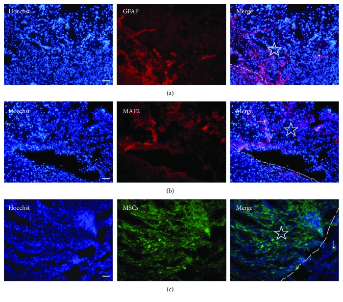
Figure 5.
Cell migrations in the PLGA scaffold in the brain with TBI. (a) Astrocytes (arrow) stained with anti-GFAP (red) migrated in the PLGA scaffold. (b) Neurons (arrow) stained with anti-MAP2 (red) migrated in the PLGA scaffold. (c) MSCs (green, arrow) migrated out of the MSC-PLGA scaffold complex. “☆” shows the PLGA scaffold or the MSC-PLGA scaffold complex in the brain, and the dashed line indicates the boundary of the scaffold. Bar = 50 μm.

