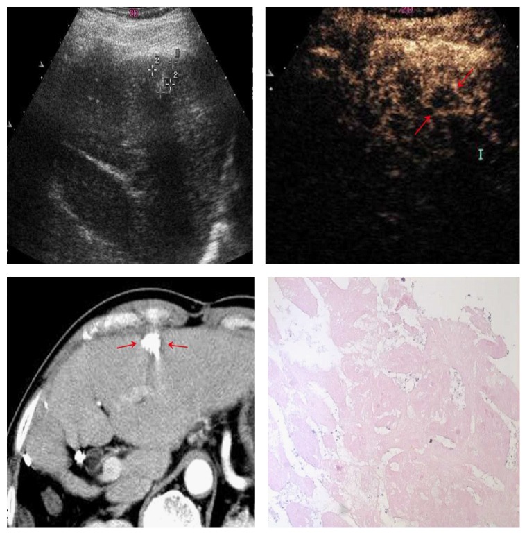
Figure 8.
35-year-old man with 1.6 cm×1.3 cm lesion (cross mark). CEUS images in the arterial phase (a, b) showed peripheral rim-like arterial phase enhancement (red arrows), probably representing reactive hyperemia, while the CT examination showed complete lack of enhancement (c) (red arrows). Pathological results showed complete necrosis of the tumor (d).

