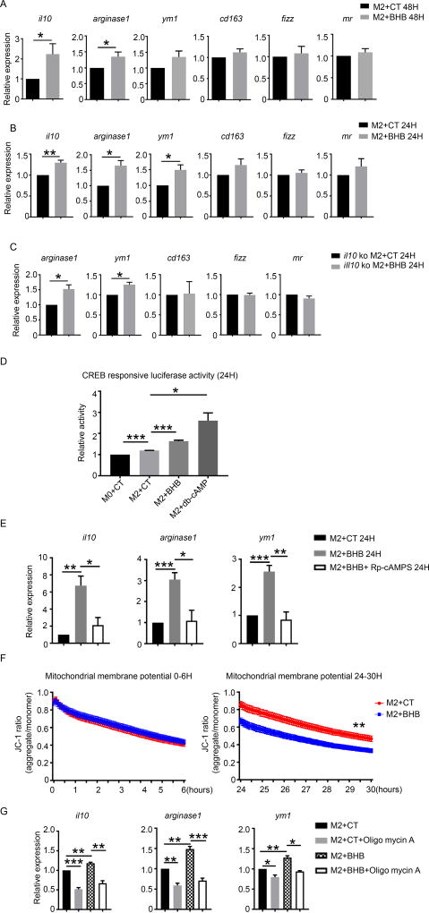
Fig. 6. BHB promoted M2 gene expression through decreasing mitochondrial membrane potential.
Bone marrow derived macrophages (BMDMs) from wild-type mice were cultured under M2 condition (IL-4 and IL-13) for 48 hours (A) and 24 hours (B) with and without BHB (15mM). BMDMs from IL-10 deficient mice were cultured under M2 condition (IL-4 and IL-13) for 24 hours (C) with and without BHB (15mM). (A and B) mRNA levels of M2 associated gene were detected in WT BMDMs. (C) mRNA levels of M2 associated gene were detected in IL-10 deficient BMDMs. (D) cAMP response element-luciferase activity in HEK cells treated under M2 condition for 24 hours with addition of control saline (CT), BHB or dbCAMP separately. (E) mRNA levels of M2 associated genes were detected WT BMDM cultured under M2 condition (IL-4 and IL-13) and BHB for 24 hours with and without Rp-cAMPS (100µM). (F) Mitochondrial membrane potentials were measured in WT BMDMs under M2 condition (IL-4 and IL-13) for 24 hours with and without BHB (15mM). (G) mRNA levels of M2 associated gene were detected in WT BMDMs treated with BHB (15mM) and oligomycin A (5µM) to maintain the mitochondrial membrane potential. Data are mean and SEM from three or four independent experiments. Data were analyzed with Student’s t-tests. * P<0.05; ** P<0.01; ***P<0.001.

