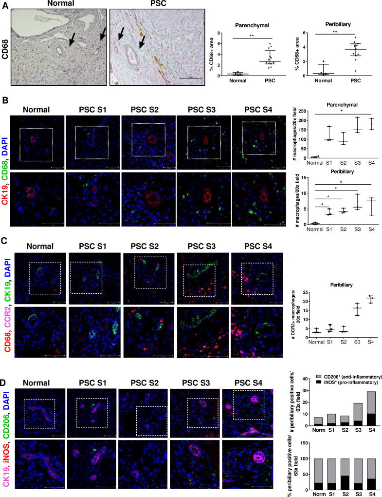
Fig. 1. Macrophages accumulate in the peribiliary areas of the livers of PSC patients.
(A) CD68 immunohistochemistry on healthy and cirrhotic-stage PSC livers (20×); quantification of CD68+ area. Arrows: bile ducts. (B) CD68 (total macrophages), CK19 (cholangiocytes) co-immunofluorescence (40×); quantification of CD68+ cells (n=4 normal, n=3 PSC). (C) CD68, CK19 and CCR2 (recruited macrophages) co-immunofluorescence (40×); quantification of CCR2+ cells (n=3/group) (D) CK19, iNOS (pro-inflammatory macrophages) and CD206 (anti-inflammatory macrophages) co-immunofluorescence (63×); quantification of iNOS+ and CD206+ cells (4 normal; 3 PSC). Higher magnifications of the dotted areas are shown in the lower row. *p<0.05; **p<0.005 (Mann-Whitney).

