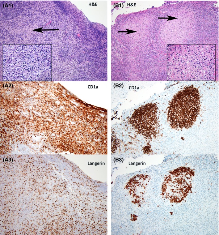
Figure 1.

Panel (A) Florid dermatopathic lymphadenopathy is characterized by paracortical expansion by small lymphocytes, histiocytes, and Langerhans cells (A1: H&E—Hematoxylin and Eosin, original magnification—100× and inset—400×).The reactive Langerhans cells express CD1 a (A2‐100×) and Langerin (A3‐100×). Panel (B) In contrast, Langerhans cell histiocytosis shows the infiltration of Langerhans cells restricted to the lymph node sinuses (B1: H&E—Hematoxylin and Eosin, original magnification—100× and inset—400×). The neoplastic Langerhans cells express CD1a (B2‐100×) and Langerin (B3‐100×)
