
FIGURE 2.
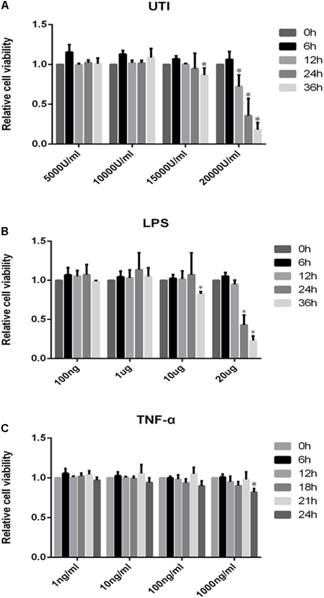
Effects of UTI, LPS, and TNF-α on viability of PMVECs. Showing cell viability of PMVECs treated with UTI (A), LPS (B), and TNF-α (C) at different doses and for different durations. In the range of 5000–10000μ/mL, UTI (A) did not affect the cells’ viability at 0–36 h; however, massive cell death occurred at 15000 U/mL of UTI at 36 h treatment (p = 0.006) and at 20000 U/mL at 12, 24, and 36 h (p = 0.002, 0.005, and 0.000, respectively). LPS (B) at concentration between 100 ng/mL and 1 μg/mL did not affect the cells’ viability at 0–36 h. When treated with LPS at 10 μg/mL for 36 h (p = 0.023) and 20 μg/mL for 24 and 36 h (p = 0.017 and 0.000), a higher incidence of cell death was detected in comparison with untreated controls. TNF-α (C) only at 1000 ng/mL could affect the cell viability at 24 h (p = 0.008). Considering the above, UTI at concentration of 10000 U/mL for 24 h, LPS 1 μg/mL for 24 h, and TNF-α 10 ng/mL for 18 h were adopted for subsequent in vitro studies. All values represent mean ± SD in triplicate; ∗represent significant differences, compared with corresponding control group, p < 0.05.