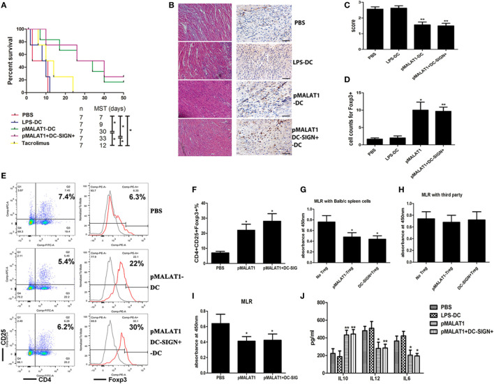
Figure 6.
Adoptive transfer of MALAT1-overexpressing dendritic cells (DCs) protected mice from acute transplant rejection and induced Tregs expression. Transplant-recipient mice were transfused with phosphate-buffered saline (PBS), LPS-stimulated DCs, MALAT1-overexpressing DCs (pMALAT1-DCs), or sorted DC-SIGN+ cells from MALAT1-overexpressing DCs (pMALAT1+DC-SIGN+) before transplantation or oral treatment with 1 mg/kg/day of tacrolimus for 7 days. (A) Graft survival times of recipients. Abbreviation: MST, median survival time. The survival times in the groups were compared using Mann–Whitney U testing. Transfusion with MALAT1-overexpressing DCs or DC-SIGN+ DC populations significantly prolonged cardiac allograft survival compared with LPS-DC and PBS transfusion. (B) Histologic studies of cardiac allografts harvested 7 days after transplantation were stained with hematoxylin and eosin (H&E) (left) and immunohistochemically stained for Foxp3 (right) [(B), original magnification 20×]. (C) Assessment of H&E staining by grading according to the 2005 classification of the International Society for Heart and Lung Transplantation for acute cellular rejection. (D) Cell counts of infiltrating Foxp3+ cells in cardiac allografts from each group 7 days post-transplantation by immunohistochemical staining. Cardiac allografts from recipients transfused with MALAT1-overexpressing DCs and DC-SIGN+ subpopulations had more Foxp3-positive staining cells than did those from recipients transfused with LPS-DC and PBS. The data indicate mean ± SD values derived from five samples in each group and were compared using analysis of variance with the Ryan method. (E,F) Tregs (CD4+CD25+ Foxp3+) in splenic T cells were assessed by flow cytometry (E) and are shown as percentages (F). Treg numbers were significantly elevated in the spleens of recipient mice injected with pMALAT1-DCs or pMALAT1-DC-SIGN+ DCs. Filled histograms represent isotype-matched irrelevant specificity controls. (G,H) Tregs (CD4+CD25+) isolated from recipients transfused with pMALAT1-conditioned DCs by MACS were added to cocultures with mitomycin C-treated splenic cells (from BALB/c mice or third-party mice) and T cells. The suppressive ability of Tregs was assessed by T cell proliferation using BrdU-ELISA. Tregs significantly decreased T cell proliferation when cocultured with donor splenic cells, but not when cocultured with third-party splenic cells. (I) Splenic T cells were separated from recipient mice at day 7 post-transplantation. The proliferating activity of splenic T cells was assessed by BrdU-ELISA. In the recipients transfused with pMALAT1-DCs or pMALAT1-DC-SIGN+ DCs, splenic T cell proliferation was significantly suppressed. (J) IL10, IL12, and IL6 concentrations in the sera of recipient mice were measured by ELISA. The IL12 and IL6 in serum were significantly decreased and IL10 was significantly increased in mice transfused with pMALAT1-DCs or pMALAT1-DC-SIGN+ DCs. The data are presented as the mean ± SD from at least five independent experiments. *P < 0.05; **P < 0.01. pMALAT1-DCs, MALAT1-overexpressing DCs; pMALAT1+DC-SIGN+, DC-SIGN+ subsets from MALAT1-overexpressing DCs.

