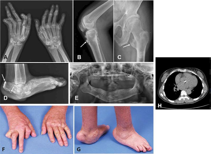
Figure 2.
Radiologic and clinical features of patient 2 of family 1938. A-D, Radiographs of the hands (A), left knee (B), left femur and hip (C), and left foot (D) at age 45 years, demonstrating significant deformities and ectopic calcification at the sites of tendon insertions (arrows). E, Radiograph of the jaw, demonstrating a complete absence of teeth. F and G, Photographs of the hands (F) and feet (G) at age 45 years, demonstrating musculoskeletal deformities. H, Transverse computed tomography image of the chest, demonstrating dense ectopic calcification of the aortic valve. Color figure can be viewed in the online issue, which is available at http://onlinelibrary.wiley.com/doi/10.1002/art.40179/abstract.

