Figure 5.
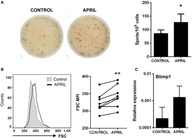
APRIL increases the number of IgM-secreting cells. Splenocytes were incubated with APRIL (1 μg/ml) or media alone for 48 h and then plated in ELISPOT plates previously coated with anti-IgM mAb, for a further 24 h. After incubation, cells were washed away and a biotinylated anti-trout IgM mAb used to detect number of spot forming cells. (A) Images from a representative experiment are shown together with a quantification of spot-forming cells, as mean + SD (n = 7). (B) After 72 h of stimulation with APRIL, splenocytes were labeled with anti-trout IgM mAb and analyzed by flow cytometry. IgM+ B cells were gated and the mean fluorescence intensity (MFI) for their forward scatter (FSC) determined. Representative histogram and FSC MFI values for different individual fish under control or APRIL conditions are shown (n = 7). (C) Splenocyte cultures were treated with APRIL (1 µg/ml) or left unstimulated for 24 h, and then RNA from IgM+ FACS isolated B cells was extracted as described in Section “Materials and Methods.” The transcription of Blimp-1 relative to the endogenous control EF-1α was calculated for each sample, and shown as mean + SD. Asterisks denote significantly different values in APRIL-treated cultures compared with control cultures (*p ≤ 0.05 and **p ≤ 0.01).

