
Figure 1.
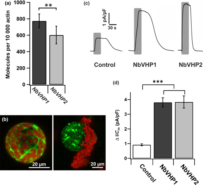
Expression and function of NbVHPs in Nicotiana benthamiana mesophyll cells. (a) Transcript abundances of NbVHP1 and NbVHP2 in mesophyll protoplasts isolated from noninfiltrated leaves. n = 4 independent experiments, means ± SE; **, P < 0.01, Student's t‐test. (b) Confocal fluorescence images of a protoplast (left) and vacuole (right) released from different mesophyll cells overexpressing NbVHP1 together with free green fluorescent protein (GFP). Red fluorescence is a result of chloroplast autofluorescence. Bars, 20 μm. (c) Representative pyrophosphate‐induced current responses of vacuoles released from mesophyll cells overexpressing free GFP together with either NbVHP1 or NbVHP2 after agroinfiltration. As a control, GFP was overexpressed alone. Duration of pyrophosphate treatment (150 μM) is indicated by the superimposed gray bars. Total number of experiments with individual vacuoles is given in (d). (d) Maximal pyrophosphate‐induced changes in current density of vacuoles released from mesophyll cells overexpressing free GFP alone (control, n = 10) or free GFP together with either NbVHP1 (n = 14) or NbVHP2 (n = 4) after agroinfiltration. Pyrophosphate was applied at a concentration of 150 μM. Data points represent means ± SE. Asterisks indicate significant differences compared with the control (Student's t‐test): ***, P < 0.001.