
Figure 3.
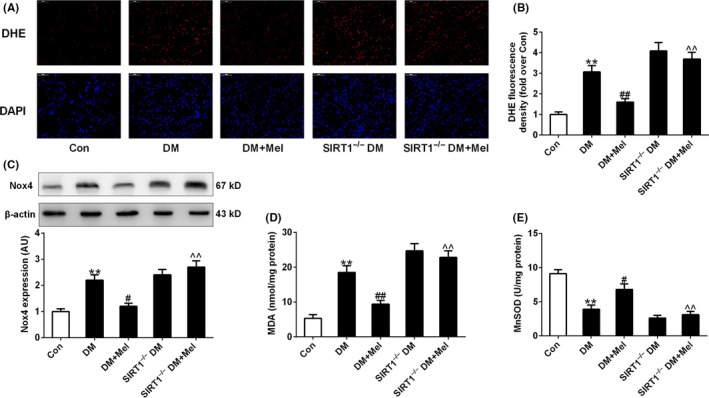
Melatonin inhibited oxidative stress in diabetic hearts but not in SIRT1−/− diabetic hearts. (A) Representative microphotographs of DHE staining in heart sections. Original magnification × 400. (B) Quantitative analysis of DHE fluorescence density (fold over Con). (C) Protein expression of Nox4. (D) Myocardial malondialdehyde (MDA) content. (E) Mitochondrial manganese superoxide dismutase (MnSOD) activity. Presented values are means ± SEM. DM, diabetes mellitus; Mel, melatonin. n = 8 in each group. **P < .01 vs Con. # P < .05, ## P < .01 vs DM. ^^ P < .01 vs DM+Mel