
Figure 9.
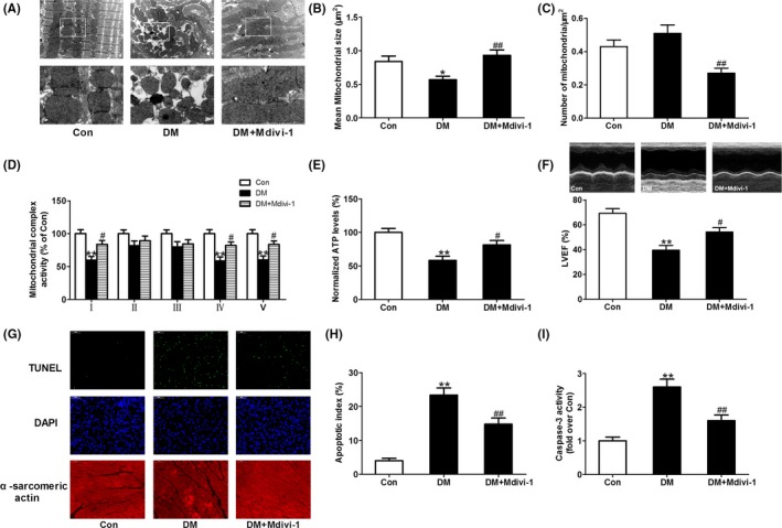
Inhibition of mitochondrial fission with mdivi‐1 improved mitochondrial function and cardiac function and reduced cardiomyocyte apoptosis in diabetic mice. (A) Representative transmission electron microscopic images of the myocardium (major finding is B,C). (B) Mean size of mitochondria. (C) The number of mitochondria per μm2. (D) Mitochondrial complex activity (percentage of Con). (E) Normalized ATP levels. (F) Left ventricular ejection fraction (LVEF). (G) Representative photomicrographs of TUNEL‐stained and DAPI‐stained heart sections. Original magnification ×400. (H) Apoptosis index. (I) Myocardial caspase‐3 activity (fold over Con). Presented values are means ± SEM. DM, diabetes mellitus. n = 8 in each group. *P < .05, **P < .01 vs Con. # P < .05, ## P < .01 vs DM