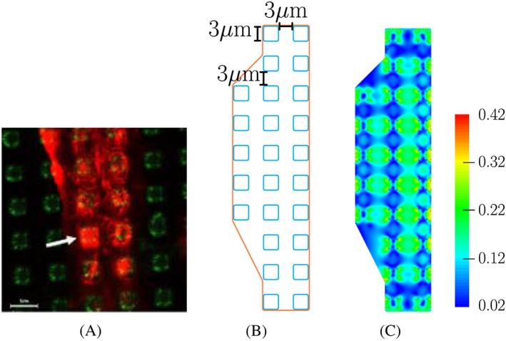
Figure 4.

Experimental and predicted distributions of actin in osteoblasts. A, Actin formation of MG‐63 osteoblasts on 3μ m×3μ m pillar array as described by Matschegewski et al.6 B, Sketch of the cell used in our simulation. C, Our predicted actin distribution characterised by the measure Π in Equation 7
