Figure 4.

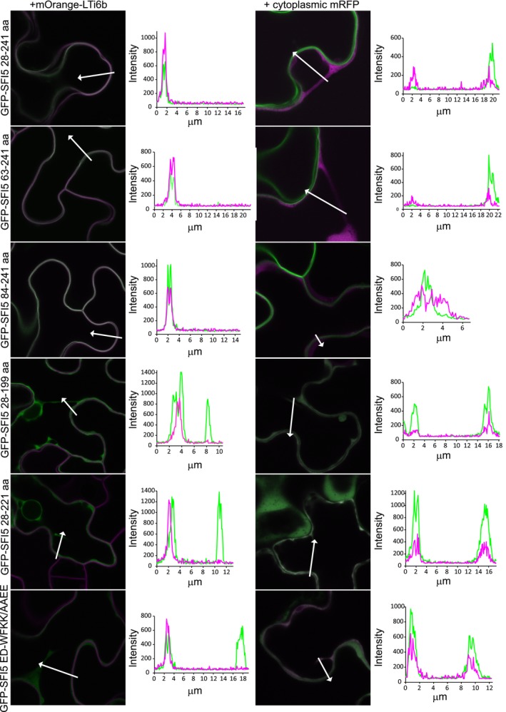
The calmodulin (CaM) binding motif of SFI5 is required for plasma membrane localization in Nicotiana benthamiana leaves. Single optical section images of GFP‐tagged SFI5 deletion and mutant forms (as indicated to the left of the images) coexpressed with the PM marker mOrange‐LTi6b or cytoplasmic mRFP (as indicated above the image columns). The fluorophores are shown in green for GFP and magenta for the mRFP or mOrange. The arrows indicate the lines drawn to generate the intensity profiles shown to the right of each image. Each profile was drawn such that both a cytoplasmic strand, or region of cytoplasm, and the plasma membrane were crossed. The lengths of the profiles are shown in the graphs in μm and the intensities are as generated by the confocal software. GFP, green fluorescent protein.
