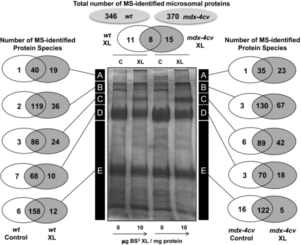
Figure 2.

Mass spectrometric identification of crosslinked muscle proteins with an altered electrophoretic mobility. Shown is a Coomassie‐stained SDS‐PAGE gel with chemically crosslinked microsomes from wild‐type (wt) versus dystrophic mdx‐4cv skeletal muscle. Lanes 1 to 4 are non‐treated wt muscle versus 10 μg BS³/mg protein‐incubated wt muscle versus non‐treated mdx‐4cv muscle versus 10 μg BS³/mg protein‐incubated mdx‐4cv muscle preparations, respectively. The number of MS‐identified proteins in the high to low molecular mass zones A‐E of the analysed gel lanes is illustrated by Venn diagrams.
