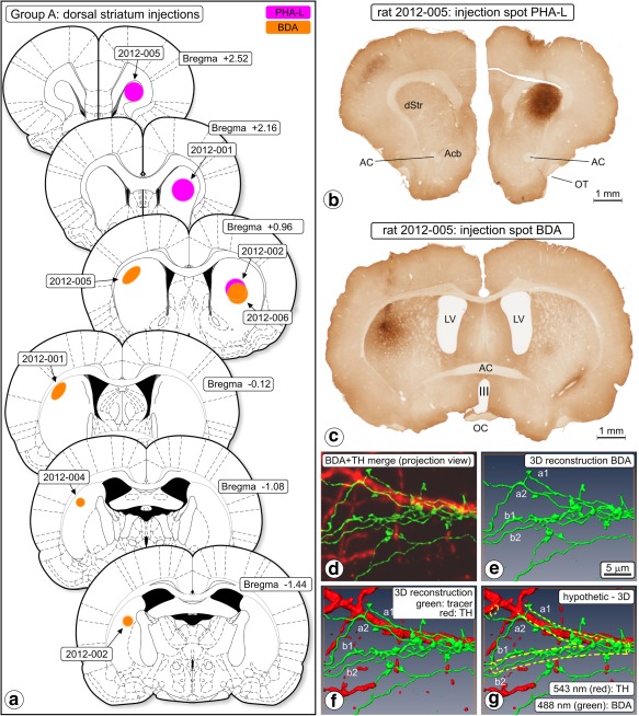
Figure 1.

Anterograde tracing, dorsal striatum injections (Group A). (a) Chartings of injection sites. (b) Photomicrograph of a section of experiment 2012‐005 showing the center of the Phaseolus vulgaris‐Leucoagglutinin (PHA‐L) injection spot. (c) In the same animal and more caudally, biotinylated dextran amine (BDA) was injected into the contralateral hemisphere. III = third ventricle; AC = anterior commissure; Acb = nucleus accumbens; LV = lateral ventricle; OT = olfactory tract. (d–g) “Woolly” fibers. BDA‐labeled fibers and their boutons in ventral tegmental area (VTA), rat 2010‐006. The “woolly” configuration here consists of two fibers (a,b) running parallel on each side of an (unstained, i.e., imaginary) dendrite, wrapping around that dendrite and forming boutons. (d) Merged projection view of the images obtained in two‐channel confocal scanning (BDA green, TH red). (e) 3D reconstruction of the BDA labeled fibers, (f) merged 3D reconstruction; BDA‐labeled fibers and TH expressing structures. (g) Hypothesized dendrite added (yellow dashed lines). Scale marker in (e) holds for all frames. “Woolly” fiber terminals were in all our observations involved with an “invisible dendrite” and never with a TH‐immunopositive dendrite [Color figure can be viewed at http://wileyonlinelibrary.com]
