Figure 2.

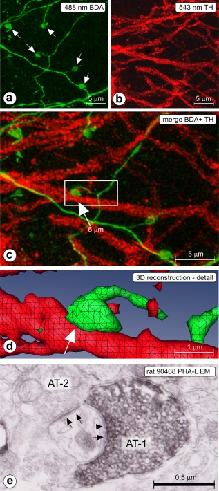
(a–d) Rat 2012‐006; CLSM sample acquired in substantia nigra pars reticulata (SNr). (a) 488 nm channel; Z projected image showing labeled, thin fibers, and boutons (arrows), (b) 543 nm channel; Z projected image showing tyrosine hydroxylase (TH) immunofluorescent dendrites. (c) Z‐projected merge image. BDA = green, TH = red. Apposition between labeled fiber and TH dendrite is indicated with an arrow. The boxed area is enlarged and presented in 3D reconstruction in (d). Apposition indicated in frames (c) and (d) with arrow. Such appositions are suggestive for the existence of a synapse. (e) Rat 90468; electron micrograph taken in SNr (injection of PHA‐L in Acb‐c, see Figure 5a), showing a synapse (arrows) between the Phaseolus vulgaris‐Leucoagglutinin‐labeled axon terminal (AT‐1) and a thin dendrite. The same dendrite receives also a synaptic contact from an axon terminal of unknown origin (AT‐2) [Color figure can be viewed at http://wileyonlinelibrary.com]
