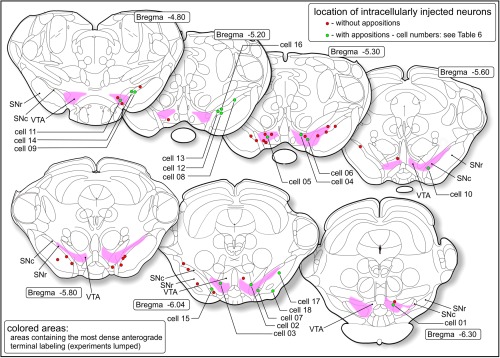
Figure 6.

Combined anterograde‐retrograde tracing/intracellular injection study (Group C). Locations of retrogradely labeled neurons in the ventral tegmental area (VTA), substantia nigra pars compacta (SNc), or substantia nigra pars reticulata (SNr) that were successfully filled with Lucifer yellow. The cells indicated with green circles had appositions with fibers labeled with anterograde tracer. Numbers of these cells correspond with those in Table 6. The colored zones correspond with the areas where the highest densities were present of labeled striatomesencephalic fibers. Around these areas labeled fibers occur much sparser [Color figure can be viewed at http://wileyonlinelibrary.com]
