
Figure 8.
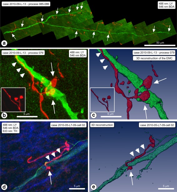
Examples of appositions of boutons of ventral striatal afferent fibers in SN on dendrites of intracellularly Lucifer yellow (LY)‐injected, dorsal striatum (dStr) projecting neurons. (a) Case 2010‐009‐L‐12 on process 085/086 (cell body not recovered). Montage of projection images covering a LY labeled dendrite (green) and the striatal fibers in the area, with appositions (arrows). (b) Same rat; LY dendrite (process nr 079). High magnification projection image of one of the appositions, a typical “earmuff” apposition (EMC). BDA = red. Inset: EMC seen in animal nr. 106. (c) 3D computer reconstruction of the EMC (arrows) shown in frame (b). Note that the BDA‐labeled fiber runs for a while alongside the dendrite (arrowheads). The inset is the 3D reconstruction of the inset of frame (b). (d) Another example of an “earmuff” apposition. Case 2010‐005. (e) 3D computer reconstruction of this EMC (arrows) of the sample shown in frame (d) [Color figure can be viewed at http://wileyonlinelibrary.com]